Soil Food Web Epi-Fluorescence Microscope Kit
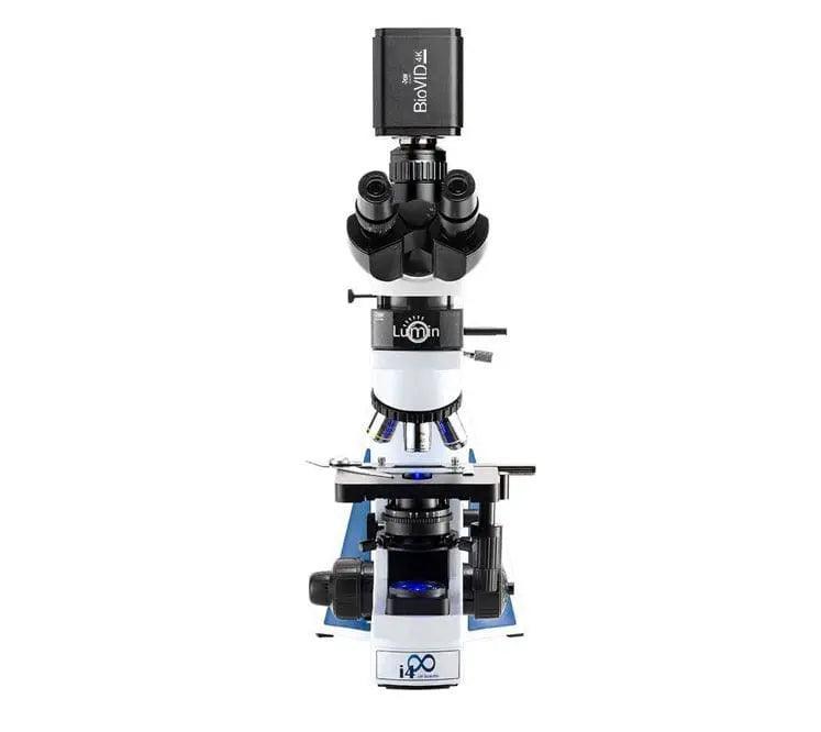
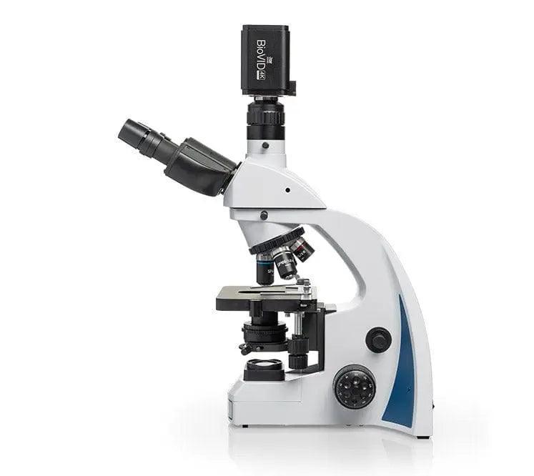
SKUI4M-TN4A-ISL3, i4P-EPiL-485N & BVC-4K16-CMT3
Product Overview
There are two distinct uses for epi-fluorescent microscopy:
First, colonization of roots by mycorrhizal fungi is easy to see (Ames, R. and E. Ingham, 1984) as VAM fungi produce autofluorescent compounds at the interface of the arbuscular cell wall and the plant cell membrane. With gridded cameras images, it is easy to see the percentage of the total root length occupied by active arbuscules colonization can be easily assessed. Count the number of grids in root that has fluorescent arbuscules present in the grid and count the total grids looked at. Grids with fluorescent arbuscules divided by total grids with and without fluorescent arbuscules times 100 gives percent colonization. Work by John Moore and a second person back in 1999 (I hope the reference for this paper is in our website list of references) showed that in grasses, 12% colonization gives measurable benefit to the plant, but 40% colonization allows ALL the benefits to be realized.
The second use for epi-fluorescent microscopy involves use of fluorescein diacetate (FDA, original paper by Bengt Soderstrom, around 1976, and should be in reference material on the website).
One application is to assess coverage of leaf surfaces, fruit, or bark by layers of organisms growing on those surfaces. Only the active organisms will take up the stain and fluoresce. Strips of the surfaces can be cut out, stained by placing the materials in fluorescein diacetate for 3 minutes, and the stain will be taken up and metabolized by active, growing cells. Placement of the leaf, bark or fruit on a slide illuminated by the fluorescein wavelengths allows the percentage of the total plant surface where active organisms cover the surface to be assessed. Coverage around 70% of higher active bacteria and fungi will result in reduction of fungal and bacterial diseases on the leaves, wood and fruit. Coverage is the important factor so that disease organisms never reach the plant surface, foods to allow germination of the spores or reproductive particles of the disease organisms will be consumed by the beneficial organisms first.
A second application of staining with FDA is to add the FDA to a dilution of soil, compost, extract or tea for a 3 minute period of time, then add hot, molten agar, which will stop all cells from taking up more FDA (The cells should be killed or at least slowed significantly). Place the sample in a well-slide, NOT a hemocytometer slide, however (see Ingham et al 1985 for description and details), place the slide on the microscope and assess number of active bacteria (stained yellow-green fluorescein color), the length of fungal hyphae stained the fluorescein color, and then assess total bacteria and total fungi using shadowing microscopy.
Vital staining of fungi in pure cultures and in soil with fluorescein diacetate. / Söderström, Bengt.
Includes:
- i4 Trinocular Microscope with Infinity Semi-Plan Objectives
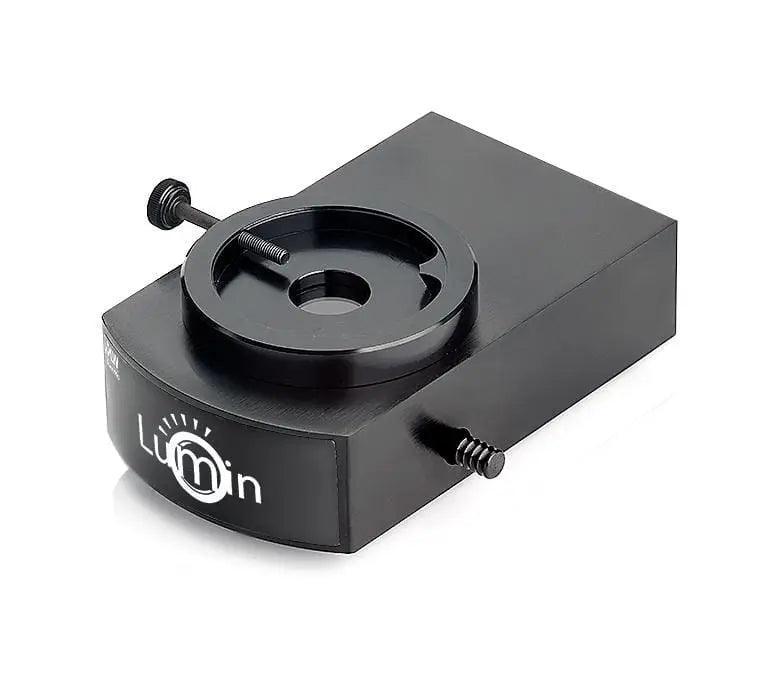
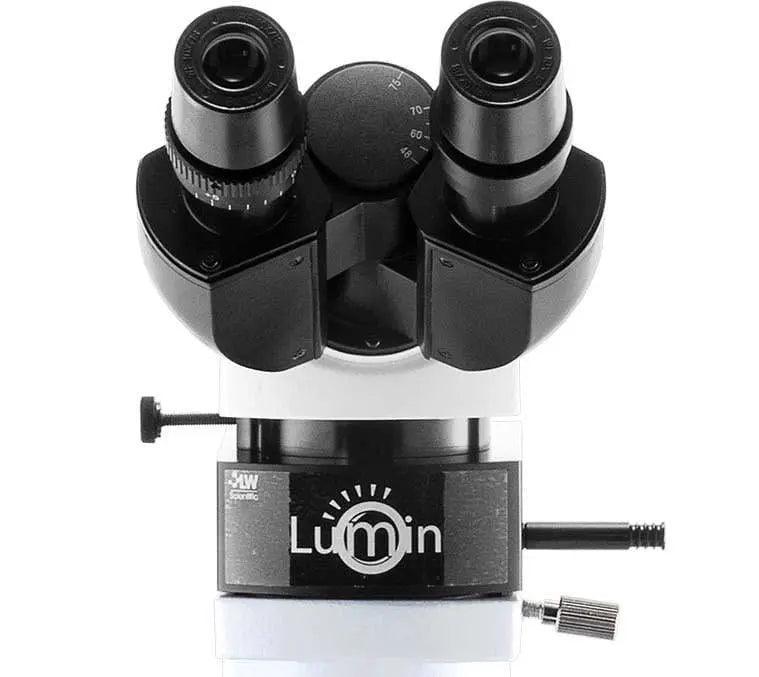
- Lumin Epi- Fluorescence Module
- BioVID HD 4K Microscope Camera
Support & Resources
Support & Resources
Couldn't load pickup availability

Soil Food Web Epi-Fluorescence Microscope Kit
Recently Viewed
Subscribe to our emails


